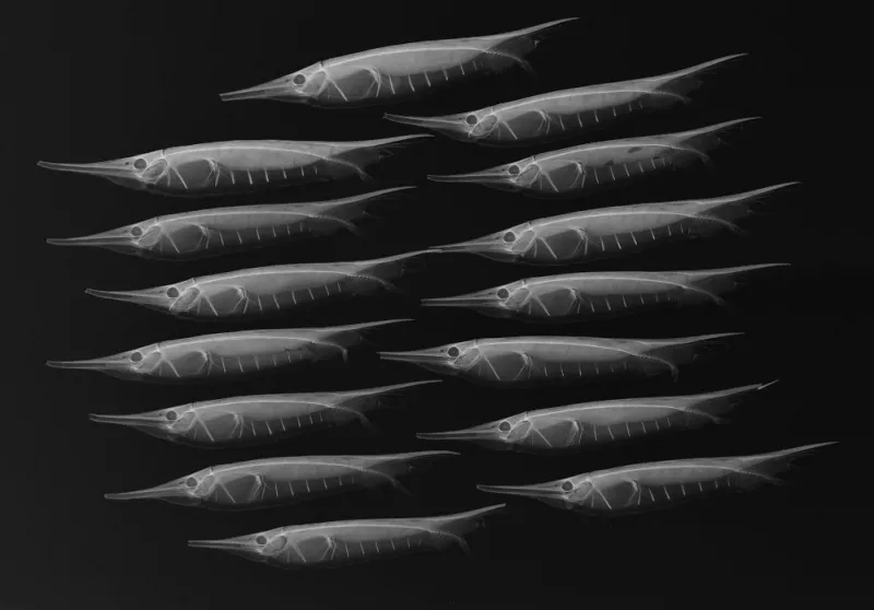
X-Ray Image of Grooved Razorfish

An X-ray image of grooved razorfish (Centriscus scutatus). Razorfish are encased in thin, transparent bony plates attached to their spines, which you can see in the X-ray. Also known as shrimpfish, razorfish have a unique swimming style: they keep their bodies vertical (heads down, tails up) while propelling themselves forward in schools. Note that the back of the fish is bony and nearly straight; all of its fins are on its belly.
Scientists in the Division of Fishes at the Smithsonian's National Museum of Natural History use X-ray images, like the one shown, to study the complex bone structure and diversity of fish without having to dissect or damage the specimen. In 2012, the National Museum of Natural History hosted "X-Ray Vision: Fish Inside Out," a temporary exhibit that showcases fish evolution and diversity through 40 black and white X-ray images prepared for research purposes. See more photos from the exhibit.

