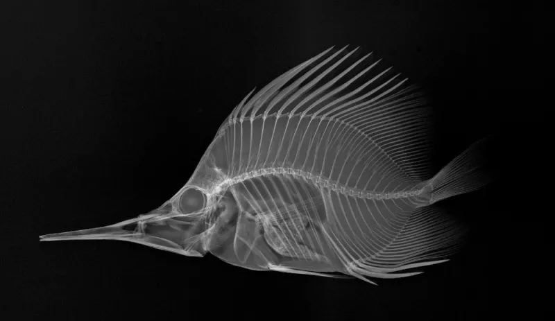
X-Ray Image of a Longnose Butterflyfish

The clearly pictured spines, rays and snout make identifying this longnose butterflyfish (Forcipiger longirostris), which was collected in French Polynesia in 2004, straightforward in this X-ray image. Like butterflies, many butterflyfish have dramatic coloration, often yellow and black, and “eyespots” on their bodies. Scientists in the Division of Fishes at the Smithsonian's National Museum of Natural History use X-ray images, like the one shown, to study the complex bone structure and diversity of fish without having to dissect or damage the specimen.
In 2012, the National Museum of Natural History hosted "X-Ray Vision: Fish Inside Out," a temporary exhibit that showcases fish evolution and diversity through 40 black and white X-ray images prepared for research purposes. See more photos from the exhibit.

