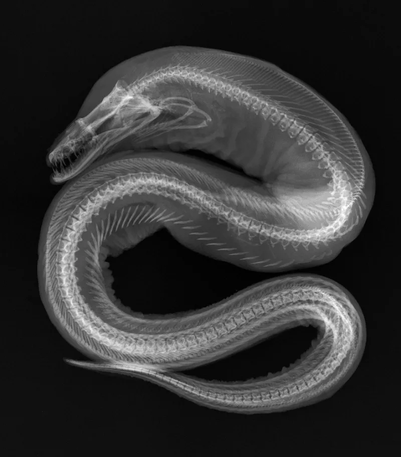
X-Ray Image of a Viper Moray Eel

In the X-ray image of this Viper Moray Eel (Enchelynassa canina), note the second set of jaws in the “throat”; these are the gill arches, which are present in all fish. Gill arches support the gills, the major respiratory organ of fish. Scientists in the Division of Fishes at the Smithsonian's National Museum of Natural History use X-ray images, like the one shown, to study the complex bone structure and diversity of fish without having to dissect or damage the specimen.
In 2012, the National Museum of Natural History hosted "X-Ray Vision: Fish Inside Out," a temporary exhibit that showcases fish evolution and diversity through 40 black and white X-ray images prepared for research purposes. See more photos from the exhibit.

