

Trials and Tribulations with Contamination

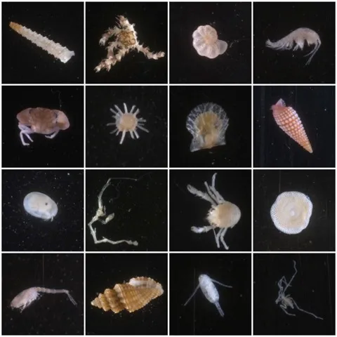
My internship at the Smithsonian National Museum of Natural History started off without a hitch. I was excited to look through the microscope at samples collected from halfway across the world in Papua New Guinea (PNG). My first job was to sort the tiny organisms in the samples. I was on the hunt for foraminifera (tiny single-celled organisms), as well as mollusks (like mussels and clams) and crustaceans (especially crabs and shrimps).
The samples came from devices called Autonomous Reef Monitoring Structures (ARMS). Each device consists of stacks of PVC plates designed with open and closed spaces to house organisms, similar to the spaces that coral reefs have. ARMS are used around the world but this specific project will help give scientists an idea of what the future ocean will look like due to impacts from ocean acidification. PNG is a prime location for this because of its vast diversity and the naturally occurring carbon dioxide (CO2) that seeps up from volcanic cracks in the seafloor. These CO2 seeps cause the surrounding water to become more acidic. Scientists are worried that the reefs in acidic water are more likely to lose biodiversity, which could affect the food chain.
The ARMS are placed on coral reefs near these seeps and on reefs about 100 meters away from the seeps where the water is not acidic. By comparing samples from the two conditions, scientists will be able to see what the future of the ocean holds as the acidity of the ocean continues to increase because of rising levels of carbon dioxide due to human activities. The ARMS that were deployed in PNG were retrieved after two years and processed in Papua New Guinea, and then the organisms were shipped to the Museum, which is where I came in.
I was familiar with most of the organisms (like crabs and mussels), but before my internship I had never even heard of foraminifera, so learning to distinguish these very small creatures was a challenge. But, after a day or two of learning the species I became accustomed to the sorting. It became so ingrained that at the end of the day I would still see the organisms each time I closed my eyes to sleep or blink.
After sorting through the animals that were found on the ARMS, we moved on to the lab. I felt confident that my experience and ability to learn quickly would make the transition easy. Boy was I wrong. To start off, the DNA from our sorted organisms had to be taken out of its home in the cells of the organisms. Once the DNA had been taken out we had to amplify our target area of DNA—meaning we made multiple copies of that area.
That was when things got challenging. While amplifying the selected area of DNA on our first trial we encountered contamination. When working with DNA this is a dreaded word to hear. In our case we could see there was contamination because the control sample came back from analysis with signs of DNA, when none should be present. This is a problem because the DNA of the single-celled foraminifera that we sorted would be overpowered if any other DNA was introduced. Since our samples originally contained lots of different kinds of organisms, including bacteria that are everywhere, and everyone in the lab works with DNA, the contamination could have come from anywhere.
To narrow down the source of contamination in our second attempt, we put the tubes and water we were using in our experiment under UV light, which destroys any DNA present. As an added precaution we also disinfected the lab table with bleach to remove any unwanted DNA, like dead skin cells that would naturally come from a person’s body or bacteria found in dust. After taking these precautions we gave it a second try and still found the dreaded contamination.
Ever heard of the saying “third time's a charm”? For us that was not the case. More like ”bad things come in threes.” Even with more precautions—moving our work under a hood that kept the area free of dust and unwanted contaminants, disinfecting everything with bleach, using new equipment, and even turning the UV light on everything to destroy any DNA left behind--our efforts came up short. With so much effort and attention to detail, we were positive our third attempt was going to work and that our troubles would be over. But the search for the source of the contamination continued over the weekend.
The next day, I got an email from my mentor saying that she had figured out why we were having a problem with contamination. Something called the “master mix” was the culprit. Since the master mix was contaminated and it went into all our samples, even the ones that were purposely left with no DNA as the control, it meant that all our amplified DNA would be contaminated. After figuring out where our problem was coming from we were able to move forward with the rest of the samples and get them all done on schedule to prepare for the next step.
Despite the obstacle of contamination, the data generated made all the frustration of dealing with it worthwhile. All the tiny organisms that I looked at under the microscope and couldn’t differentiate finally had their differences revealed by differences in their DNA. Having gone through this process from beginning to end was an amazing experience that showed me what the scientific process was really like.