

The distinctive form of a winghead shark (Eusphyra blochii) is revealed by an X-ray image. The Winghead Shark, one of about ten species of hammerhead sharks, has its eyes set at the tips of its wide, T-shaped head, giving it superb binocular vision.
slideshow
X-Rays of Fish Reveal Diversity
Scientists in the Division of Fishes at the Smithsonian's National Museum of Natural History use X-ray imaging to study the complex bone structure and diversity of fish. This image gallery showcases X-ray images of sharks, their relatives, and bony fish, revealing how some fish have skeletons built from cartilage while others are built from bone.
In 2012, the National Museum of Natural History displayed "X-Ray Vision: Fish Inside Out," a temporary exhibit that showcased fish evolution and diversity through 40 black and white X-ray images prepared for research purposes. Each X-ray is paired with a photograph of the preserved fish specimen, demostrating the value of radiography as a means of study that does not damage or destroy the specimen. See the touring schedule to find out where this exhibit will be shown next, through 2015.
To see even more photos from the exhibit, visit Encyclopedia of Life's X-Ray Vision Collection.


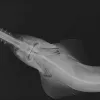


X-Ray Image of a Winghead Shark
Credit: © Sandra Raredon/Smithsonian Institution
X-Ray Image of a Slender Snipe Eel
Credit: Sandra J. Raredon / Smithsonian InstitutionThe elongated body, characteristic long and narrow snout, and small teeth make the slender snipe eel (Nemichthys scolopaceus) easily identifiable in this X-ray image. Snipe eels live at great depths in the ocean: this specimen was collected at over 2,000 meters (more than a mile down) in the North Atlantic. They swim with their mouths open, feed on invertebrates, and have over 700 vertebrae—more than any other animal.

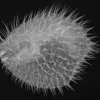
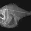
X-Ray Image of a Long-Spined Porcupine Fish
Credit: Sandra J. Raredon / Smithsonian InstitutionThe robust oval, spine covered body of a long-spined porcupine fish (Diodon holocanthus) is revealed in this X-ray image. To ward off predators, a porcupinefish inflates its body by pumping water into its stomach, transforming the fish into a round, rigid ball bristling with spines.

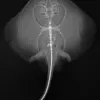
X-Ray Image of a Monterey Skate
Credit: © Sandra Raredon / Smithsonian InstitutionAn X-ray image of a Monterey skate (Raja montereyensis) reveals a spine that extends like a tail out from the pelvic fin. The skeletons of skates, rays, chimaeras, and sharks are made of cartilage, rather than bone. Scientists in the Division of Fishes at the Smithsonian's National Museum of Natural History use X-ray images, like the one shown, to study the complex bone structure and diversity of fish without having to dissect or damage the specimen.
In 2012, the National Museum of Natural History hosted "X-Ray Vision: Fish Inside Out," a temporary exhibit that showcases fish evolution and diversity through 40 black and white X-ray images prepared for research purposes. See more photos from the exhibit.

X-Ray Image of Grooved Razorfish
Credit: Sandra J. Raredon / Smithsonian InstitutionAn X-ray image of grooved razorfish (Centriscus scutatus). Razorfish are encased in thin, transparent bony plates attached to their spines, which you can see in the X-ray. Also known as shrimpfish, razorfish have a unique swimming style: they keep their bodies vertical (heads down, tails up) while propelling themselves forward in schools. Note that the back of the fish is bony and nearly straight; all of its fins are on its belly.

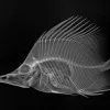
X-Ray Image of a Longnose Butterflyfish
Credit: Sandra J. Raredon / Smithsonian InstitutionThe clearly pictured spines, rays and snout make identifying this longnose butterflyfish (Forcipiger longirostris), which was collected in French Polynesia in 2004, straightforward in this X-ray image. Like butterflies, many butterflyfish have dramatic coloration, often yellow and black, and “eyespots” on their bodies.

X-Ray Image of a Smalltooth Sawfish
Credit: © Sandra Raredon/Smithsonian InstitutionThe long toothy rostrum or “saw” gives sawfish their common name. They use the saw to dig in the sand for crustaceans or to attack prey by vigorously slashing from side to side. This smalltooth sawfish (Pristis pectinata) is endangered in the U.S. because of habitat loss as mangroves are destroyed, and because their saws are easily caught in fishing nets.

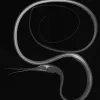
X-Ray Image of a Viper Moray Eel
Credit: © Sandra Raredon/Smithsonian InstitutionIn the X-ray image of this Viper Moray Eel (Enchelynassa canina), note the second set of jaws in the “throat”; these are the gill arches, which are present in all fish. Gill arches support the gills, the major respiratory organ of fish.

X-Ray Image of a Tropical Hatchetfish
Credit: © Sandra Raredon/Smithsonian InstitutionTropical hatchetfish (Argyropelecus lychnus), like the one shown in this X-ray photograph, live in the dark depths of the ocean; this specimen was collected at about 2,789 feet (850 meters) in the Pacific. As with many other deep-sea fishes, their eyes are large and their bellies have numerous small cream-colored spots (or “light organs”) that are bioluminescent. Each species emits its own unique pattern of light.